When people think of eye exams, they immediately think of eye charts and prescription lenses. While these tools play a valuable role in vision care, a comprehensive eye exam includes much more. The goal is to gain detailed insights into your overall eye health, and a visual field test—something many people may be unfamiliar with—is one important component.
A visual field test maps your field of vision—both central and peripheral. It creates a detailed map of everything you can see off to the side, when looking straight ahead.
This test helps us detect blind spots, areas of reduced vision, and other visual field defects that might suggest underlying eye conditions or neurological issues.
How Visual Field Tests Work
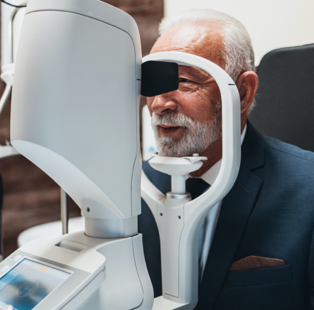
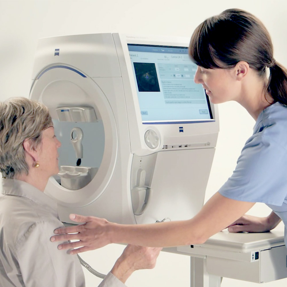
Visual field testing might sound complex, but the process is quite straightforward. During the test, you will sit in front of what’s called a perimeter. The entire process typically takes 10 to 15 minutes, depending on the type of screening and your specific needs.
Here is what happens during a typical visual field test:
You will place your chin on a rest and look straight ahead at a central target light. One eye is covered while the other is tested. Small lights of varying brightness will flash at different locations around your peripheral vision. Each time you see a light, you’re supposed to press a button.
The test itself requires concentration and patience. You must focus on the central target while staying attentive to the small lights appearing in your peripheral vision. Some patients find this challenging initially, but most adapt quickly.
There is a learning curve for this test, and it is normal to feel uncertain about whether you are seeing the dimmer lights. The machine is designed to account for normal variations in responses.
This instrument systematically tests hundreds of points across your visual field, creating a detailed map of your vision sensitivity. Areas of normal vision appear as expected patterns, while reduced sensitivity or blind spots (where you do not respond to the light stimulus)show as darker regions or gaps.
Isolated gaps might be normal variations, while consistent patterns across multiple tests suggest genuine visual field defects, which warrant a closer evaluation.
Types of Visual Field Tests
There are several types of visual field tests, each designed for specific purposes and conditions, including:
- Confrontation Visual Field Testing: A simple, quick, and preliminary visual field check that helps identify large or obvious defects. The results are basic, but they can flag potential concerns that signal the need for further evaluation.
- Automated Perimetry: This test is highly sensitive, detecting even subtle changes. We can use it to detect and monitor glaucoma, neurological diseases, or other subtle visual field changes over time.
Amsler Grid Testing: This test assesses the central visual field and is commonly used for tracking age-related macular degeneration. Irregularities may indicate damage to the macula.
What Conditions Can Visual Field Tests Detect?
Visual field testing helps us detect potential concerns at an early stage, giving us the best chance to protect your vision. It also allows us to closely monitor the progression of existing eye conditions and track how well you’re responding to treatment, so we can adjust your care as needed.
Glaucoma
Glaucoma is the most common reason for visual field testing. The disease causes characteristic patterns of vision loss, typically starting with small blind spots in the peripheral vision that gradually enlarge. The test helps determine the severity and speed of progression of glaucoma and guides treatment decisions.
Retinal Conditions
Conditions like macular degeneration, diabetic retinopathy, and retinal detachments can cause specific visual field defects. These patterns help pinpoint the affected area and guide treatment decisions.
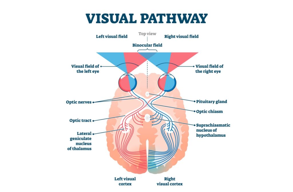
Neurological Conditions
Strokes, brain tumours, and other neurological conditions affecting the visual pathways can cause distinctive visual field patterns. For example, a stroke affecting the right side of the brain might cause left-sided vision loss in both eyes. Sometimes, visual field testing is the first indication of these serious health issues.
Optic Nerve Disorders
Conditions affecting the optic nerve, such as optic neuritis or optic nerve head drusen, can cause various visual field defects that help with diagnosis and monitoring.
When Do You Need a Visual Field Test?
Everyone can benefit from this test as part of their routine eye exam, but it is especially important for people experiencing specific symptoms or those with certain risk factors:
- You may need a visual field test if you are over 40 and have risk factors for glaucoma, such as a family history, elevated eye pressure, or belonging to certain ethnic groups.
- Patients with diabetes should have visual field testing as part of their diabetic eye exams, as diabetic retinopathy can affect peripheral vision.
- If you are experiencing symptoms such as tunnel vision, blind spots, or difficulty with side vision.
- If you are taking certain medications (i.e., hydroxychloroquine) that can affect vision or have neurological symptoms that might indicate visual pathway problems.
When we combine visual field testing with retinal images (fundus photo and retinal scans), the results provide detailed insights into your overall visual health.
Take Control of Your Eye Health
Routine eye exams are your first line of defense against sight-threatening eye conditions. Diagnostic screenings, including visual field tests and retinal scans, help us paint a full picture of your overall eye health.
If it has been a while since you last sat in an exam chair or if you have never had a comprehensive visual screening, it might be time to visit our Downtown Vision Care team. Contact us today to request an appointment.